Cardiac and vascular ultrasound
Cardiac and Vascular Ultrasound — Dr Agueznai, Cardiologist in Casablanca

Cardiac ultrasound (echocardiography) and vascular Doppler use ultrasound waves to visualise the heart, its valves and the blood vessels in real time. Dr Agueznai has been performing transthoracic and transoesophageal echocardiography for more than 12 years.

What is a cardiac and vascular ultrasound?

A cardiac ultrasound, or echocardiogram, is non-invasive and completely painless. Its purpose is to obtain images of all the structures of the heart, i.e. the valves and chambers (atria and ventricles), and to assess how well the heart muscle is contracting.

To determine whether the symptoms you are experiencing (shortness of breath and chest pain) are caused by a heart problem; to assess the impact of certain diseases on your heart, such as high blood pressure and pulmonary arterial hypertension; to diagnose a heart defect or a disease of the heart muscle (cardiomyopathy); to establish a diagnosis of heart failure; to observe or assess abnormalities of the heart valves (valvular heart disease); to check for the presence of a blood clot; and to examine the pericardium (the sac surrounding the heart) or the thoracic aorta.

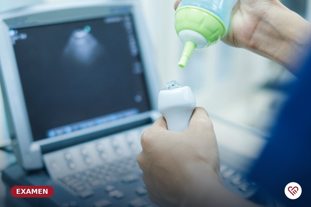
For the examination, you will be asked to undress to the waist and lie comfortably on the examination couch so that the cardiologist can apply a gel to the area to be examined. You will be asked to keep still while the ultrasound probe is moved over your chest.

To help you plan, please note that your visit may last between 30 and 45 minutes in total.

This information is provided for guidance only and does not replace a medical consultation.